Visual recognition mechanisms and hemispatial neglect
With hemispatial neglect, patients present the symptom that information from half the visual field (usually the left half) is difficult to detect, even though there is nothing wrong with the patient’s sight. However, the problem with hemispatial neglect is due not to damage to the occipital lobe, which is the gateway to visual information processing, but to damage in the frontal and parietal lobes in many cases. But why would damage to other regions, rather than damage to the occipital lobe, cause problems with visual processing?
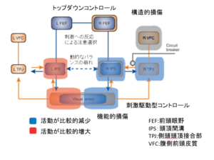
Visual information is basically processed along a pathway that runs from the occipital lobe through the temporal lobe (ventral stream: semantic information processing) or the parietal lobe (dorsal stream: spatial information processing) towards the frontal lobe, as shown in this diagram,

but in fact, in addition to the pathways from the occipital lobe to the frontal lobe (bottom-up pathways), there are more complicated pathways that run from the frontal lobe towards the temporal lobe, the parietal lobe or the occipital lobe (top-down pathways), and the region that is activated and the extent of the activation is adjusted via the top-down pathways according to the condition that the individual is placed in and various needs.

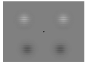
The paper I discuss today investigates what brain activity occurs relating to visual information processing in hemispatial neglect patients, using visual-evoked potential. In the study, researchers displayed a visual stimulus in one of four sections of a screen and measured how the subjects’ brains reacted differently to visual stimuli on the left or right.

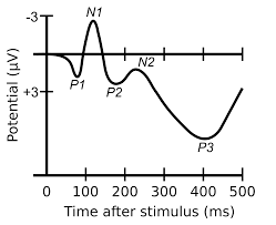
The study investigated slight changes in the brain waves over time with event-related potential and the changes in brain activity related to them,

but the results did not show a large difference in brain activity between hemispatial neglect patients and the control group at the initial stage after displaying the visual stimulus (P1: low-level visual processing), while at later stages (N1, P2), activity in the intraparietal sulcus and the striate and extrastriate areas decreased, suggesting that hemispatial neglect may be due to damage to the top-down pathways.
I thought that the visual processing system is complicated.
Reference URL: Impaired visual processing of contralesional stimuli in neglect patients: a visual-evoked potential study.
[Abstract]
Transient visual-evoked potentials (VEPs) were recorded in 11 patients with right brain damage and spatial neglect. High-resolution EEG was recorded using focal stimuli located in the four visual quadrants. VEPs to left stimuli, i.e. located in the neglected side, were compared to VEPs to right stimuli. Results showed that bottom-up processing of a visual stimulus located in the neglected hemifield was intact up to approximately 130 ms from stimulus onset. Hemispheric differences were not significant for either C1 or P1 components representing the activity of striate and extrastriate areas, respectively. In contrast, visual processing in more dorsal areas adjacent to the superior parietal lobe was changed from normal. We failed to record the N1a component for left visual field stimuli expected in the 130-160 ms time range. Furthermore, the N1p (140-180 ms) and P2 (180-220) components were delayed and/or reduced in amplitude for stimuli located on the neglected side. The source of the N1a was previously localized in the intraparietal sulcus in the dorsal occipital cortex; N1p may represent a reactivation of area V3A and P2 reactivation of occipital visual areas including V1 due to top-down feedbacks. Six patients with left brain damage (LBD) and no neglect and 21 healthy subjects were also tested in the same experimental conditions used for patients with neglect. In LBD patients, all components evoked by contralesional stimuli were comparable to ipsilesional components. Overall, data allow localizing in time and space the processing deficit specific for patients with neglect. The first takes place around 130 ms in the bottom-up processing at the level of the anatomically intact dorsal parietal areas; the second is located at the level of the reactivation of the striate and extrastriate areas via feedback connections from higher visual areas. The two functional impairments were limited to left-field stimuli.


